Breast Assessment
Australian
Breast Cancer
Statistics
Breast cancer is the most common cancer in Australian women accounting for 27.4 per cent of all new cancers.
The risk of developing breast cancer before the age of 85 in women is 1 in 8. Survival rates after diagnosis of breast cancer in women have increased in recent years.
Between the periods 1982–1987 and 2006–2010, five-year survival has increased from 72 per cent to 89.4 per cent in Australian women.
At the end of 2008, it was estimated that there were 159,325 Australian women alive who had been diagnosed with breast cancer in the previous 27 years, including 57,327 women diagnosed in the previous 5 years.
Early detection of breast cancer saves lives
Breast
Symptoms
Awareness of any subtle changes in your breast allows detection of breast cancer at an early stage. Prompt treatment can save your life. Monthly self breast examination (two weeks after your last menstrual period if you are pre-menopausal), as well as yearly breast examination with your GP is a sensible strategy to detect any subtle changes in the breasts.
Please look for the following symptoms:
– Breast lump
– Nipple discharge
– Breast pain
– Changes in skin, nipple and shape or size of the breast
Breast
Screening
There is well-established scientific evidence that participating in regular mammographic breast screening saves lives.
The target (invited) age range for breast screening has been increased from 50-69 years to 50-74 years.
Women aged 40-49 years and over 75 years are eligible for free two yearly screening mammograms even though they are not formally invited.
How to participate in breast screening – Breast Screen Australia
High Risk Groups
Familial
Breast Cancer
About 5% of breast cancers can be explained by an inherited gene mutation, most commonly in BRCA1 and BRCA2 genes. As our understanding of genetic basis of breast cancer is rapidly increasing, more forms of familial breast cancers are being described.
If you have multiple family members spanning multiple generations with breast and /or ovarian cancer, your family may be carrying a gene mutation. The risk is also increased if the family members had breast cancer at young age, cancers affecting both breasts or there are male relatives with breast cancer.
Calculate your risk of breast cancer – Cancer Australia 2014
Previous
Radiation Therapy
to the chest
Those who had received radiotherapy to the chest for conditions like lymphoma at a young age are also at risk of developing breast cancer after several decades of the treatment.
If you think that you belong to these high-risk groups please contact your GP who can refer you to a breast surgeon. Your breast surgeon can organize your assessment at a familial cancer clinic if appropriate. For the high risk patients there are number of effective strategies to reduce their risk of developing breast cancer or to detect them early.
Breast Assessment
Once you are referred to a Specialist Breast Surgeon the assessment consists of a number of steps.
Clinical history:
Thorough collection of information about symptoms, family history and other risk factors and general health status
Clinical examination:
Examination of breast and lymph node regions (axilla or armpit and neck)
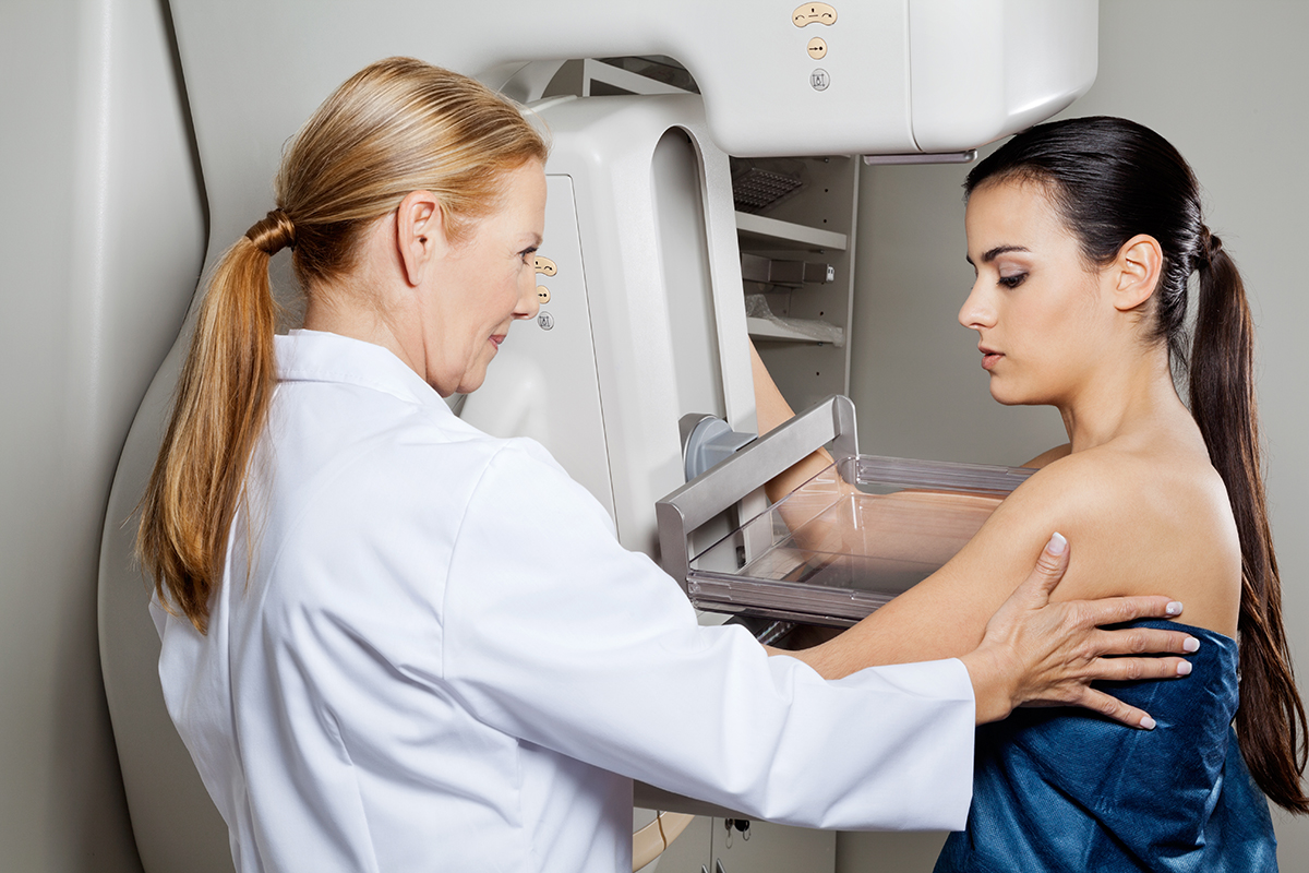
Mammography:
A Mammogram can show number of signs that may indicates breast cancer:
– Mass/density (lump)
– Suspicious collection of calcium deposits
– Differences in overall architecture of glandular elements of the breasts Some times additional special mammographic views are required to further check those abnormalities. Your breast surgeon can organize these investigations.
Breast Ultrasound:
Breast ultrasound compliments mammography and in women with dense glandular breasts (some pre- menopausal women and post menopausal women on HRT) it helps to diagnose lumps that are not clear on mammograms.
Breast MRI:
Breast MRI is a relatively new technique, which can be sometimes helpful in selected patients such as high-risk patients, those with high clinical suspicion with normal mammogram and ultrasound results or some types of breast cancers. Breast MRI is best organized by your breast surgeon after discussing its usefulness including pros and cons with you.
Breast biopsies:
Once a breast lump is found and characterized, biopsies are used to confirm or rule out cancer and other non-cancerous conditions. They are performed by specialist radiologists, usually using ultrasound as guidance. Either a very thin needle i.e. Fine Needle Aspiration (FNA) or larger needle i.e. Core Biopsy are used. However, if the abnormality is only visible on a mammogram a special mammographic biopsy named Mammotome needs to be done.
For more information Contact Us

